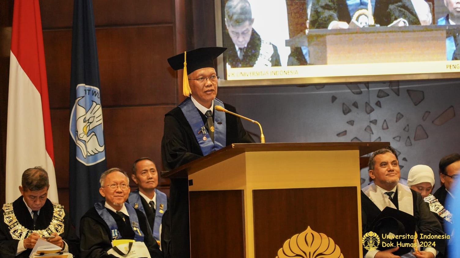
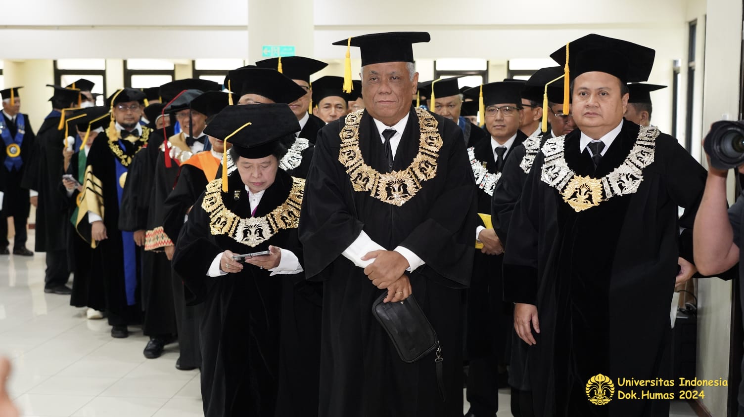
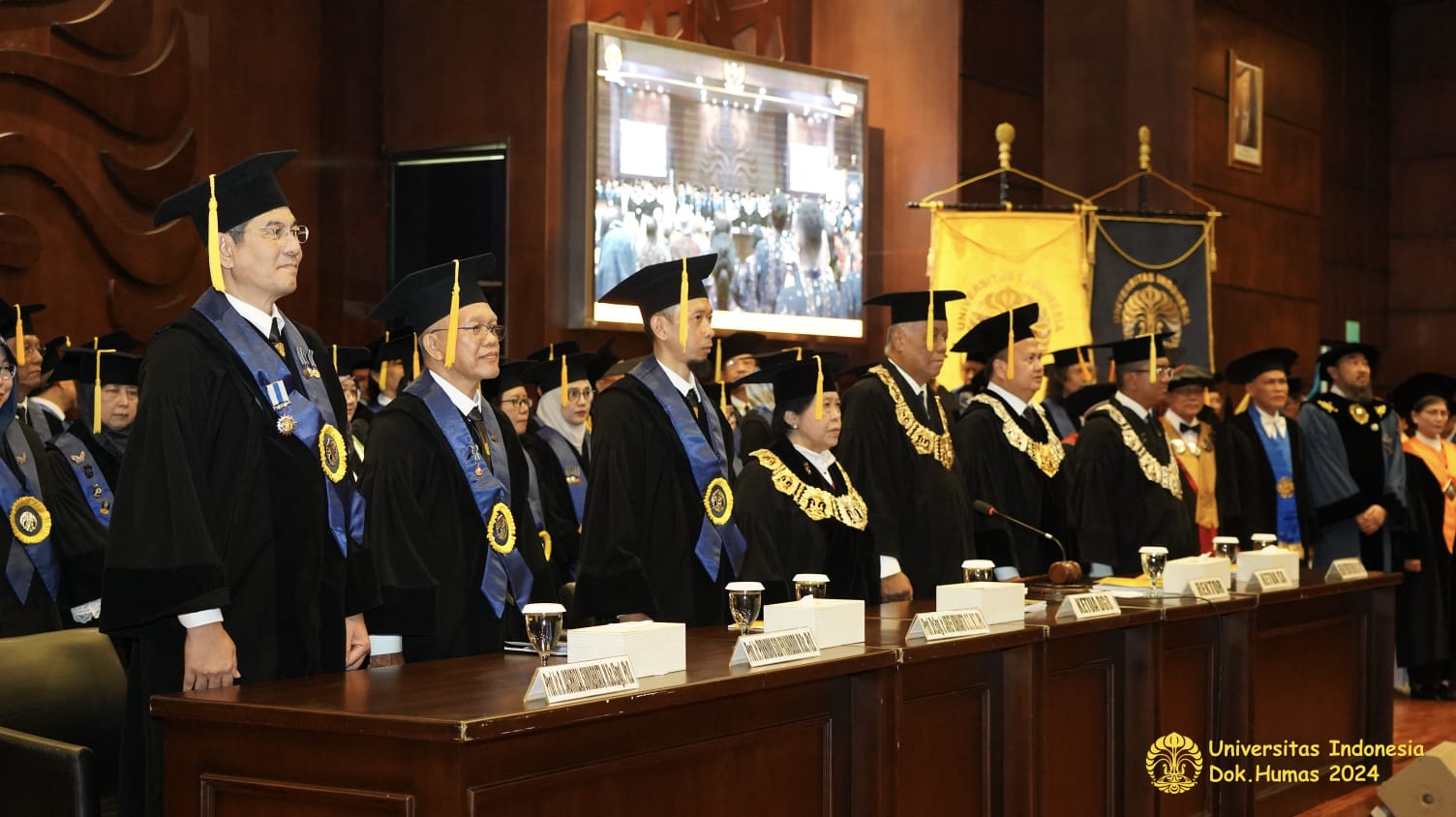
He noted that current medical diagnostic tools, such as X-rays, CT scans, SPECT, PET, and MRI, rely on high-energy media that, in excessive doses, may pose health risks. “Today’s advanced high-resolution medical imaging instruments are costly and rely on high-energy media. While these provide high-quality transmittance images due to their deep penetration capabilities, they all carry health risks,” Prof. Purnomo explained.
In response, he and his team designed an infrared tomography technique based on a matrix inverse method that is safer and energy-efficient, using infrared or near-infrared (NIR) media, which operate at lower energy levels. He described how this technology not only has sufficient penetration capabilities for the human body but is also safer than X-ray technology. “Infrared media is relatively safe and requires less energy for medical instruments, making the technology available on the market, allowing for more affordable products,” he said.
Another advantage of this technology, Prof. Purnomo added, is its ability to decompose images into constituent layers of biological tissue. Current X-ray technology only produces a combined image of all biological tissues. “In the future, the ability to decompose biological images into constituent tissue images will assist doctors in diagnosing diseases that affect only specific tissues,” he noted.
This innovation enables the visualization of each tissue layer’s thickness through image decomposition captured with infrared cameras. “This image decomposition method is expected to provide healthcare workers with an easier way to visualize the thickness distribution of each biological layer within a unified biological object,” Prof. Purnomo stated.
He also mentioned that machine learning is being optimized in the decomposition process to enhance the accuracy of images captured within the far-infrared wavelength. The technology is planned for further development to achieve 3D image decomposition, which will offer even greater benefits for in-depth medical analysis.
“We have developed an infrared tomography technique that is safe, health-friendly, portable, energy-efficient, and economical. This technology holds great promise for future medical analysis. Besides requiring expertise in photonics, this technology’s development will involve various fields, such as imaging, image processing, machine learning, and medical expertise,” Prof. Purnomo stated.
The Dean of FTUI, Prof. Dr. Ir. Heri Hermansyah, S.T., M.Eng., IPU, expressed, “The appointment of Prof. Purnomo Sidi Priambodo as a Professor in Biomedical Optoelectrotechnology at FTUI is both an achievement and a source of pride, with immense hope for the future of medical technology in Indonesia. His infrared tomography innovation is a visionary step toward providing safer, energy-efficient diagnostic solutions. This technology prioritizes patient safety while also offering energy efficiency and affordable access. At FTUI, we hope this development continues to advance through interdisciplinary collaboration to make a meaningful impact on the medical field.”
Prof. Purnomo’s research has received recognition in numerous national and international journals, with recent publications titled Decomposition Technique for Bio-Transmittance Imaging Based on Attenuation Coefficient Matrix Inverse (2024) and Image Decomposition Technique Based on Near-Infrared Transmission (2022).
Having completed his undergraduate studies in Electrical Engineering at UGM in 1987, he earned his M.Sc. from Oklahoma State University in 1996 and his Ph.D. from the University of Texas at Arlington in 2003, remaining dedicated to delivering innovative biomedical technology for Indonesia and beyond.
***
Office of Public Communication
Faculty of Engineering Universitas Indonesia


